Volume : 2 | Issue : 2
Review
Anthropometric landmarks of the mandible in a Colombian cadaveric sample
Diego Mauricio Barreto Suárez,1,2 Juan Pablo Gnecco,1 Jaime Castro-Núñez,3 Andrés Gómez-Delgado,3 Luis Fernando Gamboa,1 Germán Ayala,4 Álvaro Rodríguez-Sáenz5
1Professor, Oral and Maxillofacial Surgery Residency Program, Universidad El Bosque, Colombia
2Staff Surgeon, Oral and Maxillofacial Surgery Department, Hospital Simón Bolívar, Colombia
3Oral and Maxillofacial Surgeon, Universidad El Bosque, Colombia
4Instituto Nacional de Medicina Legal y Ciencias Forenses, Colombia
5Resident, Oral and Maxillofacial Surgery, Universidad El Bosque, Colombia
Received: February 22, 2019 | Published: April 17, 2019
Abstract
Purpose: The aim of this investigation was to obtain objective measurements of the mandible in an adult Colombian cadaveric sample and to describe their relationship to gender, age, and body mass.
Patients and Methods: A retrospective descriptive study design was implemented. To be included in the study each cadaver had to be considered legally adult and have less than 3 days of permanence at Instituto Nacional de Medicina Legaly CienciasForensesin Bogota, Colombia. Exclusion criteria were cadavers under 18 years and with any kind of bone invasive surgery in the maxillofacial area, evident bone loss, growth or craniofacial morphology alteration due to trauma or pathology, presence of exostosis or osteosynthesis material in the mandible, and subjects with syndromic abnormalities. Using a Leibinger Castroviejo caliper (Kalamazoo, MI), 2 standardized examiners measured the study variables. The information was collected between June 2006 and July 2010.
Results: A total of 250 adult Colombian cadavers were initially considered. At the end 222 cadavers (192 males and 30 females) were dissected to analyze the anatomy and anthropometry of the mandible. The studied sample had a mean age of 24 years and racially they were of Caucasian predominance and mestizo ancestral pattern. All variables measured presented significant differences between gender, except condylar heights and the central alveolar crest height. The values of the left and right mandibular ramus width presented the major differences between men and women.
Conclusion: This research objectively established the measurements of the mandible in a vast adult Colombian cadaveric sample.
Keywords: anthropometry, colombian sample, mandible
Introduction
The vocable "anthropometry" is derived from the Greek words anthropos, which signifies man, and metron, literally meaning to measure. Currently, anthropometry provides researchers the single most uncomplicated, economical, convenient, non-invasive, and universally applicable instrument for assessing the size and proportions of the human body. As such, it can be considered an invaluable technique for guiding surgeons in the diagnosis and treatment plan of many conditions affecting the craniomaxillofacial complex.
Although the outcomes of anthropometry can be applied to any human being, due to racial and ethnical differences, surgeons must be cautious when incorporating these findings into clinical practice.1 Studies, therefore, should ideally be customized to each population in order to have accurate figures which, in turn, produce better clinical results.
Given the limited amount of anthropometric studies in the Hispanic American population, and more specifically in Colombia, surgeons must rely on North American investigations. Due to the aforementioned ethnical variances, this situation represents an evident impasse that is usually neglected. The purpose of this investigation was to obtain objective measurements of the mandible in an adult Colombian cadaveric sample and to describe their relationship to gender, age, and body mass.
Material and methods
Study design and sample
A retrospective descriptive study design was designed and implemented. To be included in the study each cadaver had to be considered legally adult and have less than 3 days of permanence at Instituto Nacional de Medicina Legal y Ciencias Forensesin Bogota, Colombia and due to the retrospective nature of this study, it was granted an exemption in writing IRB. Exclusion criteria were cadavers under 18 years and with any kind of bone invasive surgery in the maxillofacial area, evident bone loss, growth or craniofacial morphology alteration due to trauma or pathology, presence of exostosis or osteosynthesis material in the mandible, and subjects with syndromic abnormalities.
Study variables
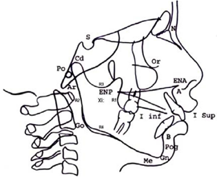
An initial cephalogram (Figure 1) was prepared in order to guide the data collection process, establishing the conventional points condilion (Co), articulare (Ar), gonion (Go), mentón(Me), gnation (Gn), pogonion (Pog), and supramentale (B). The following complementary points were assessed:
R1: The most posterior point of the mandibular ramus concavity.
R2: Projection of R1 in the posterior edge of the mandibular ramus, parallel to Frankfort plane.
R3: The most inferior point of the sigmoid notch.
R4: Projection of R3 in the inferior edge of the mandible, perpendicular to Frankfort plane.
Inferior Mandibular Plane (IMP): linear plane from gonion to menton.
Superior Mandibular Plane (SMP): linear plane from gonion to gnation.
The study active variables were the mandibular linear and angular dimensions, as follow:
Linear measurements
- Left Condylar Height (LCH): A line from condilion (Co) to its intersection to other line that crosses from R3perpendicular to the posterior border of the mandible in the left side.
- Right Condylar Height (RCH): A line from condilion (Co) to its intersection to other line that crosses from R3 perpendicular to the posterior border of the mandible in the right side.
- Left Mandibular Body Length (LMBL): Linear distance between gonion (Go) and gnation (Gn) in the left side.
- Right Mandibular Body Length (RMBL): Linear distance between gonion (Go) and gnation (Gn) in the right side.
- Left Ramus Height (LRH): Linear distance between condilion (Co) and gonion (Go) inthe left side.
- Right Ramus Height (RRH): Linear distance between condilion (Co) and gonion (Go) in the right side.
- Parasymphysiary Transversal Distance (PTD): Distance between a point consisting of the intersection between the mandibular plane (Go-Me) and a perpendicular line thatcrosses the mental foramen in the left side, and the same point in the right side.
- Intergonial Transversal Distance (IGTD): Linear distance between left gonion and right gonion.
- Intercondylar Transversal Distance (ICTD): Linear distance between left condilion and right condilion.
- Central Alveolar Crest Height (CACH): Distance between menton (Me) and the alveolar crest edge at the medial sagital line level.
- Left Alveolar Crest Height (LACH): Distance between the inferior border of the mandible and the alveolar crest edge at the first molar buccal groove level on the leftside.
- Right Alveolar Crest Height (RACH): Distance between the inferior border of the mandible and the alveolar crest edge at the first molar buccal groove level on the right side.
- Left Mandibular Ramus Width (LMRW): Distance between points R1 and R2 on the left side.
- Right Mandibular Ramus Width (RMRW): Distance between points R1 and R2 on the right side.
- Left Mandibular Body Thickness (LMBT): Distance between the most lateral point ofthe externalmandibular cortical and the most medial point of the internal mandibular cortical at retromolar trigone on the left side.
- Right Mandibular Body Thickness (RMBT): Distance between the most lateral point ofthe external mandibular cortical and the most medial point of the internal mandibularcortical at retromolar trigone on the right side.
Angular measurements
- Left Goniac Angle (LGA): An angle formed by the tangential line to the posterior border of the mandible and the mandibular plane (Go-Me) on the left side.
- Right Goniac Angle (RGA): An angle formed by the tangential line to the posterior border of the mandible and the mandibular plane (Go-Me) on the right side.
- Left Parasymphysiary Angle (LPA): An angle formed by the tangential line to the anterior basal border of the parasymphysis and the tangential line to the lateral basal border of the mandible border on the left side.
- Right Parasymphysiary Angle (RPA): An angle formed by the tangential line to the anterior basal border of the parasymphysis and the tangential line to the lateral basal border of the mandible border on the right side.
- Other variables collected and analyzed were Body Height (BH), measured in centimeters; Body Weight (BW), measured in kilograms, and the Body Mass Index (BMI), which is the subject's body mass divided by the square of the height, with the value expressed in units of kg/m2.2
Data collection
Data was collected between June 2006 and July 2010. In order to collect the information, 2 standarized examiners used a Leibinger Castroviejo caliper 0-40 mm (Leibinger, Kalamazoo, MI) for the linear measures and a goniometer for the angular variables. The intra-examiner standarization was performed measuring 20 different variables in 5 human dry mandibles donated by the Instituto Nacional de Medicina Legal y Ciencias Forensesat Bogota, Colombia in two different dates. A third examiner compared the results in a 5-day interval. The calibration was assessed by a 0-1 reliability scale using statistical analyses with SPSS 14.0 software (SPSS Inc, Chicago, Il). All values above 0.9 were considered acceptable (Table 1). The inter-examiner standardization was not implemented due to the lack of a gold standard as a reference pattern.
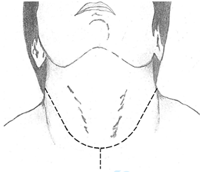
The process of data collection started with the Duque´s facial lifting technique for orofacialnecropsy.3 This method consists in a deep incision using a number 22 blade describing a Y line that starts 3 to 4 centimeters under the mastoidapophysis and moving downward to the medial line in a curvilinear way to the supra clavicular zone, then advancing vertically until the sternoclavicular joint is reached. Another curvilinear incision is performed contralaterally until joining the incision in the midline (Figure 2).
After the Y incision is performed, the procedure continues with the musculocutaneous lifting following the disection in a caudocephalic direction up to the masseter muscle, dissecting its inferior insertion and all the muscular insertions of the mentalis, depresorlabii inferioris, masseter and all insertion tissues of the internal mandibular cortical, cutting off the pterygoid and oral floor muscles, reaching the higher point of the mandibular ramus. After this, a section of the inferior insertion of the temporalis muscle tendons over the coronoid apophysis is performed, followed by the section of the articular capsule and all articular ligaments involved. The procedure ends with the complete dislocation of the mandible in order to proceed with the measuring process.
Data management and analysis
Anthropometric landmarks were computed with the Microsoft Excel® Software. The age was expressed following the 1980 WHO classification. Average values and standard deviation were computed for descriptive analysis. Height values were expressed following the 1957 Martin & Saller classification. Absolute and relative frequencies were applied for descriptive analysis.
Body mass index (BMI) was expressed following the 1980 WHO classification. Descriptive statistic was used to calculate averages, standard deviations, and confidence intervals for all mandibular landmarks. Chronological age, absolute, and relative frequencies were calculated for the height and BMI variables. All statistical data were processed with STATA 10.0 software (StataCorp LP, College Station, TX).
Results
Between June 2006 and July 2010 a total of 222 cadavers were dissected to analyze the anatomy and anthropometry of the mandible. According to (Table 2), the studied sample had a mean age of 24 years and racially they were of Caucasian predominance and mestizo ancestral pattern. 192 subjects (86.4%) were men and 30 (13.6%) were women. The average age was 24.0±3.8 years. 68.47% of subjects were classified as hipsisomes or tall (68.47%).
The most part of the active variables presented a standard deviation lower than 2.7 according with the SD stipulated for the study. All variables presented significant difference between gender, except LCH, RCH, and CACH. The values of the LMRW and RMRW presented the major differences between men and women (Table 3).
|
Examiner 1 |
Examiner 2 |
Left condylar height (LCH) |
0.997 |
0.985 |
Right condylar height (RCH) |
0.995 |
1.000 |
Left mandibular body length (LMBL) |
1.000 |
1.000 |
Right mandibular body length (RMBL) |
1.000 |
1.000 |
Left ramus height (LRH) |
0.996 |
0.999 |
Right ramus height (LRH) |
1.000 |
0.999 |
Parasymphysiary transversal distance (PTD) |
0.998 |
0.998 |
Intergonial transversal distance (IGTD) |
0.995 |
0.996 |
Intercondylar transversal distance (ICTD) |
0.999 |
0.998 |
Central alveolar crest height (CACH) |
0.988 |
0.988 |
Left alveolar crest height (LACH) |
0.978 |
0.966 |
Right alveolar crest height (RACH) |
0.992 |
0.966 |
Left mandibular ramus width (LMRW) |
1.000 |
0.999 |
Right mandibular ramus width (RMRW) |
1.000 |
1.000 |
Left mandibular body thickness (LMBT) |
0.993 |
0.986 |
Right mandibular body thickness (RMBT) |
0.993 |
0.993 |
Left goniac angle (LGA) |
0.994 |
0.984 |
Right goniac angle (RGA) |
0.987 |
0.999 |
Left parasymphysiary ANGLE (LPA) |
0.992 |
0.993 |
Right parasymphysiary angle (RPA) |
1.000 |
0.990 |
Table 1 Intraexaminer Standardization, SPSS Program 14.0. Reliability Analysis Scale.
Variable |
Males |
Females |
Total |
Number of subjects |
192 (86.4%) |
30 (13.6%) |
222 (100%) |
Mean age |
23.9±3.6 |
24.7±4.8 |
24.0±3.8 |
Height |
12/222 (5.4%) |
1/222 (0.4%) |
13/222 (5.8%) |
BMI |
0/222 (0.0%) |
0/222 (0.0%) |
0/222 (0.0%) |
Table 2 Descriptive Statistics of Total Sample.
|
Mean |
SD |
CI |
P Value |
Left condylar height (LCH) * |
17.33 |
1.95 |
16.60-18.00 |
0.09 |
Left condylar height (LCH) ** |
16.73 |
1.79 |
16.40-16.90 |
|
Right condylar height (RCH) * |
17.10 |
2.16 |
16.29-17.91 |
0.30 |
Right condylar height (RCH) ** |
16.72 |
1.80 |
16.47-16.98 |
|
Left mandibular body length (LMBL) * |
75.37 |
1.18 |
74.92-75.81 |
0.00 |
Left mandibular body length (LMBL) ** |
78.44 |
2.57 |
78.08-78.81 |
|
Right mandibular body length (RMBL) * |
75.29 |
-937 |
74.93-75.64 |
0.00 |
Right mandibular body length (RMBL) ** |
78.46 |
2.58 |
78.09-78.83 |
|
Left ramus height (LRH) * |
56.93 |
4.68 |
55.81-58.92 |
0.00 |
Left ramus height (LRH) ** |
60.33 |
2.55 |
59.97-60.69 |
|
Right ramus height (LRH) * |
57.36 |
4.16 |
55.81-58.92 |
0.00 |
Right ramus height (LRH) ** |
60.41 |
2.57 |
60.04-60.77 |
|
Parasymphysiary transversal distance (PTD) * |
35.11 |
1.03 |
34.72-35.49 |
0.00 |
Parasymphysiary transversal distance (PTD) ** |
37.67 |
2.59 |
37.30-38.04 |
|
Intergonial transversal distance (IGTD) * |
94.8 |
3.98 |
93.31-96.28 |
0.83 |
Intergonial transversal distance (IGTD) ** |
94.69 |
2.43 |
94.34-95.03 |
|
Intercondylar transversal distance (ICTD) * |
95.64 |
17.77 |
89.28-101.99 |
0.723 |
Intercondylar transversal distance (ICTD) ** |
94.7 |
2.43 |
94.35-95.04 |
|
Central alveolar crest height (CACH) * |
119.36 |
2.78 |
118.32-120.40 |
0.00 |
Central alveolar crest height (CACH) ** |
124.26 |
5.03 |
123.54-124.98 |
|
Left alveolar crest height (LACH) * |
119.65 |
2.38 |
118.75-120-54 |
0.00 |
Left alveolar crest height (LACH) ** |
124.55 |
4.92 |
123.85-125.25 |
|
Right alveolar crest height (RACH) * |
133.9 |
6.59 |
131.43-136.36 |
0.00 |
Right alveolar crest height (RACH) ** |
137.26 |
3.56 |
136.55-137.77 |
|
Left mandibular ramus width (LMRW) * |
134.06 |
5.53 |
131.99-136.13 |
0.00 |
Left mandibular ramus width (LMRW) ** |
137.17 |
3.47 |
136.68-137.77 |
|
Right mandibular ramus width (RMRW) * |
30.57 |
2.37 |
29.68-31.45 |
0.25 |
Right mandibular ramus width (RMRW) ** |
30.89 |
1.23 |
30.71-31.06 |
|
Left mandibular body thickness (LMBT) * |
26.06 |
1.60 |
25.46-26-66 |
0.00 |
Left mandibular body thickness (LMBT) ** |
27.96 |
1.09 |
27.81-28.12 |
|
Right mandibular body thickness (RMBT) * |
25.06 |
1.36 |
24.55-25.57 |
0.00 |
Right mandibular body thickness (RMBT) ** |
27.87 |
1.14 |
27.71-28.04 |
|
Left goniac angle (LGA) * |
14.09 |
-815 |
13.78-14.39 |
0.00 |
Left goniac angle (LGA) ** |
34.10 |
1.97 |
33.82-34.38 |
|
Right goniac angle (RGA) * |
14.08 |
-804 |
13.78-14.39 |
0.00 |
Right goniac angle (RGA) ** |
34.27 |
1.90 |
33.99-34.54 |
|
Left parasymphysiary angle (LPA) * |
18.11 |
3.44 |
16.82-19.39 |
0.00 |
Left parasymphysiary angle (LPA) ** |
15.44 |
-945 |
15.30-15.57 |
|
Right parasymphysiary angle (RPA) * |
17.67 |
2.45 |
16.76-18.59 |
0.00 |
Right parasymphysiary angle (RPA) ** |
15.45 |
-964 |
15.31-15.59 |
|
Table 3 Mean, Standard Deviation (SD) and Confidence Interval of Active Variables by Gender. *Females (n=30), ** Males (n=192).
Discussion
The purpose of this research was to obtain objective measurements of the mandible in an adult Colombian cadaveric sample and to describe their relationship to gender, age, and body mass. In order to achieve said goals, we analyzed 222 mandibles of human adult Colombian cadavers at the Instituto Nacional de Medicina Legal y Ciencias Forenses at Bogota, Colombia.
We studied and analyzed 222 Colombian cadavers of which 192 were men (86.4%) and 30 were women (13.6%). The average age was 24.0±3.8 years. The majority of subjects were classified as hipsisomes (68.47%) and inside the normal range of weight (61.26%), being the variables camaesome (5.8%) and underweight (0.0%) the less common. The most part of the active variables presented a standard deviation lower than 2.7 according with the SD stipulated for the study. All variables presented significant difference between gender, except LCH, RCH, and CACH. The values of the LMRW and RMRW presented the major differences between men and women.
The importance of anthropometry as a method to assess the size, proportions, and composition of the human body is unquestionable. In 1995 the WHO established the principles of anthropometry's interpretation due the interest on its applications in order to identify social an economic inequity, predict benefit, and evaluate responses during interventions. Its uses, nevertheless, need to consider different properties of the most appropriate indicators, which may variate according to individuals and populations.1
The implications of anthropometry in surgery were not recognized until recent decades,4-6 with the vast majority of the research performed in the United States of America and Europe. Currently, there is a lack of information on mandibular anthropometry in the Hispanic American population and facultative in this part of the world have to rely on foreign studies. To the best of our knowledge, this is the first anthropometric study that evaluates mandibular anthropometry on cadavers in an effort to provide objective parameters that can be utilized in craniomaxillofacial surgery.
A similar anthropometric investigation was performed by Farkas et al.7-10 in people under 18 years to analyze growth patterns. Our cadaveric sample included adult subjects who were out of the scope of the influence of major growing changes and therefore we cannot compare results. Using a different methodology, in 2004 Flórez Méndez studied 40 Peruvian female patients in order to standardize facial anthropometry and proportion with surgical implications.
Conclusion
The sample was composed of 192 (86.4%) men and 30 (13.6%) females, with a mean age of 23.9 and 24.7 years, respectively. Men were generally taller than women. Hipsisome constitutes the variable most frequently found (67.7% in males and 73.3% in females). We found significant differences in all variables between genders, being male mandibles bigger than female ones. Only two exceptions were found: Condylar heights and the central alveolar crest height. The values of the left and right mandibular ramus width presented the major differences between men and women.
The cadaveric sample we studied constitutes the major strength of the present investigation and the results can be of great value in the diagnose, treatment plan, and management of several maxillofacial conditions.
Acknowledgements
The authors would like to thank the administrative personnel at Instituto Nacional deMedicina Legal y Ciencias Forenses in Bogota, Colombia and former oral and maxillofacial surgery residents Efraín Crespo, María Carolina Gil, Mahamed Ali Gómez, Iván Torres, José Luis Claros, Jennifer Azuaje, Magda Sofía Gómez, and David Enrique Castro for their tireless efforts towards the realization of this project.
Disclosures
The authors report no conflicts of interest related to this study
References
- Physical Status: the use and interpretation of anthropometry. WHO Technical Report Series. Geneva Switzerland: World Health Organization. 1995;854.
- BMI Classification. Global Database on Body Mass Index. World Health Organization, 2006. Retrieved July 21, 2012.
- Duque AM, Sánchez E. Design of an orofacial autopsy protocol applying the facial lifting technique: Pilot study. Thesis. Pontifical Xavierian University. Bogotá, 2001.
- Farkas LG, Posnick JC, Hreczko TM, et al. Anthropometric growth study of the ear. Cleft Palate Craniofac J. 1992;29(4):324–329.
- Farkas LG, Posnick JC. Growth and development of regional units in the head and face Based on anthropometric measurements. Cleft Palate Craniofac J. 1992;29(4):301–302.
- Flórez Méndez M, Hernández I, Rossano G. Structuring and standardization of facial anthropometry according to proportions. International Journal of Cosmetic Medicine and Surgery. 2004;6:10.
- Farkas LG, Posnick JC, Hreczcko TM, et al. Growth patterns in the orbital region: A morphometric study. Cleft Palate Craniofac J. 1992;29(4):315–318.
- Farkas LG, Posnick JC, Hreczcko TM, et al. Growth patterns of the nasolabial region: A morphometric study. Cleft Palate Craniofac J. 1992;29(4):318–324.
- Farkas LG, Posnick JC, Hreczcko TM. Anthropometric growth study of the head. Cleft Palate Craniofac J. 1992;29(4):303.
- Farkas LG, Posnick JC, Hreczcko TM. Growth patterns of the face: a morphometric study. Cleft Palate Craniofac J. 1992;29(4):308–315.

