Volume : 5 | Issue : 1
Case Report
Flapless computer-guided implant surgery in the aesthetic region of a patient with lateral incisor agenesis: A case report
Matteo Miegge,1 Curci Giuseppe,2 Galati Fabiana3
1DDS, Oral surgery specialist
2DDS, Aestethic dentistry
3DDS, Orthognathodontics specialist
Received: December 23, 2022 | Published: December> 31, 2022
Introduction
Implant-prosthetic rehabilitation is the gold standard solution to rehabilitate a patient with tooth element loss in the aesthetic region.
Often, in patients with agenesis of the lateral incisor, the space available for implant fixture placement is very narrow, making implant surgery very difficult and increasing the risk of damage to the roots of adjacent teeth.
Case Report
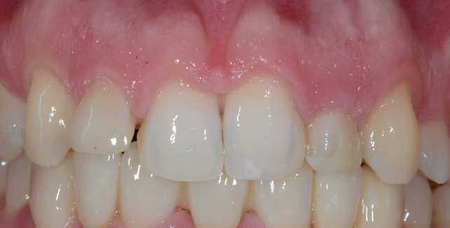
Without anamnestic comorbidities, a patient of 25 years old required a dental examination complaining of mobility of deciduous element 52.
Orthodontic therapy with splinting of the element 52 was performed at school age.
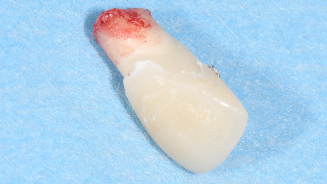
Clinically, the deciduous tooth had grade III mobility, confirmed by radiographic examination that shows considerable root resorption, the cause of the instability.
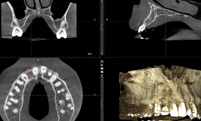
A CBCT (Computer Tac Cone Beam) was then performed to assess the surgical spaces, which were found to be very narrow:
Thickness in the buccal-palatal direction was 4.78 mm, and width in the mesiodistal direction was 4.37 mm
Due to this small bone size, a procedure with computer-guided surgery was chosen.
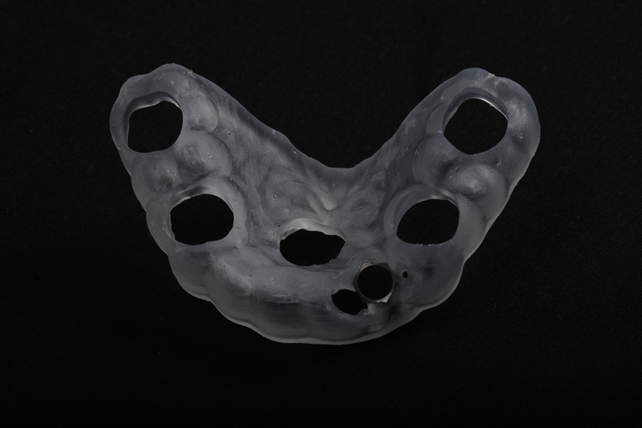
An intraoral scan was then taken to design the tooth-supported surgical template (manufactured by “JDentalcare Lab”).
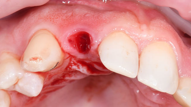
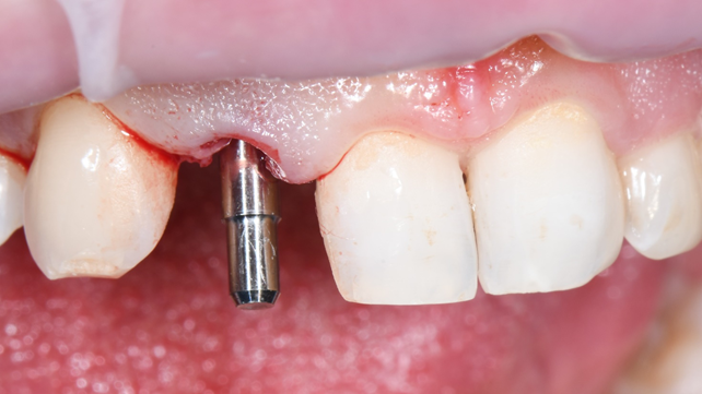
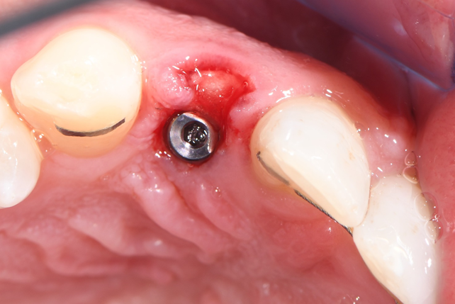
Under local anesthesia, the deciduous tooth was extracted under local anesthesia, and a JD Evolution S 3,2 x 13 mm (JDentalcare) implant was placed with preparation drills and implant placement guided by the tooth-supported surgical template. No flap for the procedure was performed; a slow-resorbing collagen sponge was inserted from the buccal side to allow adequate soft tissue support.1-3
The implant was inserted with a torque greater than 40 newton/cm, which allows immediate loading.
An intraoral scan with scan-body was taken, and after 6 hours the temporary immediately loaded element 12 crown HIPC (high impact polymer composite) was placed.
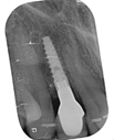
The postoperative radiograph showed correct implant placement with root preservation of dental elements 11 and 13.
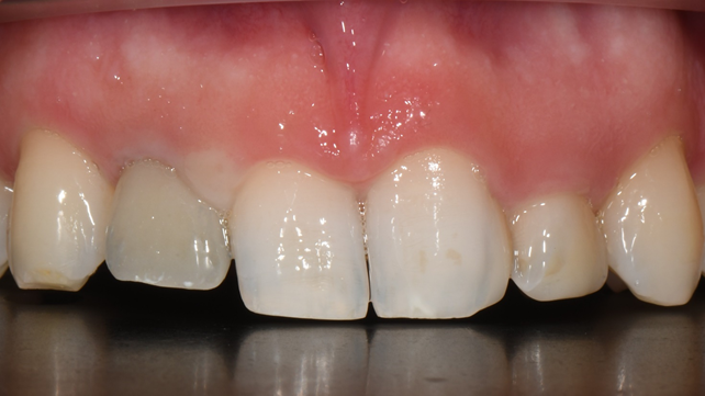
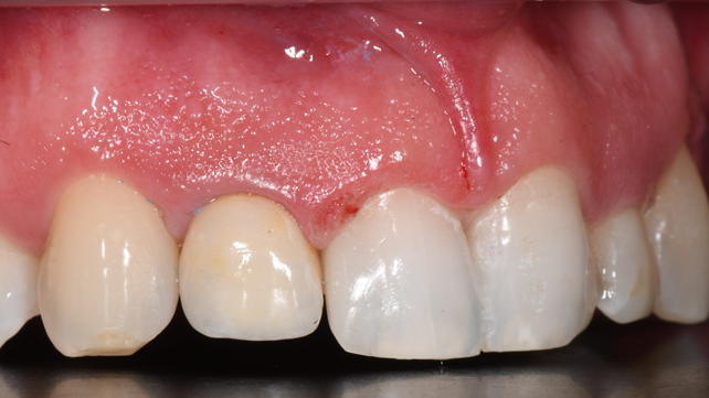
After 3 months, a new intra-oral scan was performed and then the final prosthesis of element 12 was made with a metal-ceramic crown. A metal-ceramic crown was chosen because the trans-mucosal pathway was too narrow for a zirconia crown (too thin to provide load resistance).
Before screwing in the final zirconia crown, an additive coronoplasty was performed on element 1.1 with a direct conservative composite.
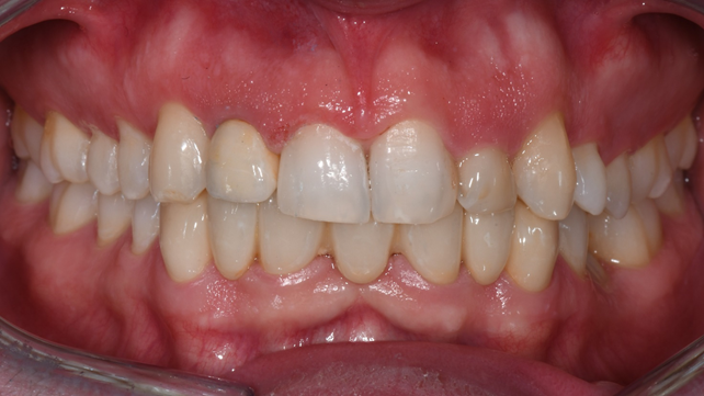
All the prosthetic elements have been manufactured by the dental laboratory “Dentalibra” (Figure 1-12).
Discussion/Conclusion
Flapless implantology allows more conservative and less painful surgery, with excellent soft tissue management in the aesthetic area.
Immediate loading allows adequate gingival tissue support while preserving apical resorption.
The tooth-supported surgical template appears to be an excellent technique for the correct placement of dental implants when there is a reduced amount of bone in the mesiodistal direction, preserving the vitality of adjacent elements.
Acknowledgements
None.
Conflicts of Interest
None.
References
- Erika Oliveira de Almeida, Eduardo Piza Pellizzer, Marcelo Coelho Goiatto, et al. Computer-guided surgery in implantology: review of basic concepts. J Craniofac Surg. 2010 Nov;21(6):1917‒1921.
- Azari A, Nikzad S. Flapless implant surgery: review of the literature and report of 2 cases with computer-guided surgical approach. J Oral Maxillofac Surg. 2008 May;66(5):1015-1021.
- Bergendal B. When should we extract deciduous teeth and place implants in young individuals with tooth. Agenesis? J Oral Rehabil. 2008 Jan;35 Suppl1:55‒63.
