Volume : 6 | Issue : 1
Case Report
Dental management of a patient with trisomy of the long arm chromosome 1: Rare case report
Mariana Gabrielli,1 Alessandra Vaz Pinto Hapner,2 Patricia Marcoccia de Souza,3 Liliane Roskamp,4 Natanael Henrique Ribeiro Mattos,5 Cintia Mussi Milani6
1DDS, Former Graduation student of Dentistry, Universidade Tuiuti do Paraná, Brazil
2DDS, Professor of Pediatric Dentistry, Universidade Tuiuti do Paraná, Brazil
3DDS, Professor of Restorative Dentistry, Universidade Tuiuti do Paraná, Brazil
4,5DDS, PhD, Professor of Endodontics, Universidade Tuiuti do Paraná, Brazil
6DDS, PhD, Professor of Stomatology and Oral Surgery, Universidade Tuiuti do Paraná, Brazil
Received: February 07, 2023 | Published: February 17, 2023
Abstract
The trisomy of the long arm chromosome 1 is a very rare Syndrome that can present specific oral manifestations such as micrognathia, high and/or narrow palate, with cleft palate in some cases. This study aimed to present a case report of the oral aesthetic rehabilitation of an 8-year-old male child diagnosed with the trisomy of the long arm chromosome 1 that had minimal school social life due to the bullying caused by his aesthetic appearance. The presence of diastema and alterations in the dental shape that caused great aesthetic disorder to the patient was corrected with the execution of direct restorations in composite resin. This simple treatment proved to be efficient to return the patient´s self-esteem and provide resocialization.
Keywords: Chromosome, Dental treatment, Oral manifestations, Syndrome, Trisomy
Introduction
A chromosome is a package of deoxyribonucleic acid (DNA) sequences found in the nucleus of the cells, composed by genes. Humans have 23 pairs of chromosomes, 22 pairs of numbered chromosomes, called autosomes, and one pair of sex chromosomes, XX in females, XY in males. Each parent contributes with one strip to each pair of chromosomes so that the offspring gets half of its chromosomes from the mother and half from the father.1-3 Each chromosome has a short arm (p) and a long arm (q), which contain the genetic information. About 10% of the human genome is characterized by chromosome 1.1,2 Duplication of the long arm of chromosome 1 is a rare abnormality, with only 39 cases reported in the world literature. It occurs when there is an extra copy of genetic material in its long arm.1-6
The intensity of signs and symptoms vary accordingly to the size and location of the genes involved.1,2 People with small duplications, near the tip of the long arm (q), are slightly affected, while those with larger duplications, which extend near the center of the chromosome, carry severe birth defects, associated with a low life expectancy.1-3 In general, the excess of genetic material in one of the 46 chromosomes increases the risk of delayed development and growth, as well as greater susceptibility to recurrent birth defects.2 Thus, the severity of duplication is closely related to which genetic material was duplicated or whether any material was lost or replicated on a chromosome in another region.1-4
Specific characteristics in patients with chromosome 1 long arm duplication syndrome include the presence of micrognathia, high and/or narrow palate, with cleft palate in some cases, mild to severe impairment in development and learning, congenital cardiac abnormalities, and short stature.1 Although the signs and symptoms are characteristic among patients with this syndrome, the literature points out that their intensity and severity may vary broadly.3
The present study aimed to review the literature of the Trisomy Syndrome of the long arm chromosome 1 to clarify the association of clinical signs and symptoms, as well as to present a case report on the oral aesthetic rehabilitation of a pediatric patient, to improve his self-esteem and resocialization.
Case Report
An 8-year-old male patient arrived with his mother at the Dental Clinic complaining about his aesthetics dental condition. The mother reported his school social life was minimal, due to the bullying caused by his aesthetic appearance. She also mentioned that, at the moment, he was not under medical monitoring, but that he had annual appointments with a neurologist since early childhood, due to seizures. He was a child with fragile health since the neonatal period, receiving the diagnosis of Microduplication Syndrome 1q44 or Trisomy of the long arm of chromosome 1, after 4 years of clinical and laboratory research.
The mother reported that the child started having problems in the first hours after childbirth. He was intubated for 18 days in the neonatal intensive care unit due to perinatal anoxia. Next, he developed persistent pulmonary hypertension, early neonatal infection, right-side pneumothorax, and noise induced seizures. Because of these seizures he wore a specific type of glasses and earplugs to reduce them. The seizures were controlled by the age of six, and nowadays he uses Oxcarbazepine 300mg and lamotrigine 25mg, every 12 hours, daily.
As for his oral development, the mother reported his primary teeth had a pointed shape and they were all spread over the dental arch. As the child presented oligodontia, along with many skin changes as cracked lips and periorbital skin, sparse eyebrows and nocturnal sudoresis since early childhood, and episodes of fever without a diagnosis, the pediatrician suggested the diagnosis of ectodermal dysplasia and referred him for a genetic investigation. The child was submitted to a postnatal screening of the entire genome to identify submicroscopic chromosomal changes by Single Nucleotide Polymorphisms-Array (SNP-array). The examination showed the presence of two submicroscopic genomic variants, both mapped on the long arm of chromosome 1 in 1q44, in close segments, but not in contiguous segments. The laboratory suggested genetic research of both parents, using the Fluorescence in Situ Hybridization (FISH) method. It was discovered that both parents presented microduplication in the 1q44 region and demonstrated that the presence of genomic variants in the parents was inherited by the child.
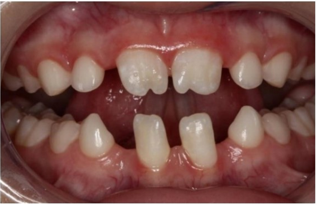
The extraoral physical examination revealed a small face, prominent ears, pallor with areas of deep dark circles around the eyes and micrognathism (Figure 1). The intraoral examination showed the permanent maxillary right and left central incisors displaying a chamfering in the incisal region, gyro version of the permanent mandibular left and right canines and permanent mandibular right central incisor and large diastema in the region of the permanent mandibular central incisors, due to the absence of the lateral ones (Figure 2). Permanent maxillary and mandibular central incisors showed changes in surface, color, texture, and translucency and a class II malocclusion was observed.

|
Figure 1 Extraoral view showing a small face, prominent ears, pallor with areas of deep dark circles around the eyes and micrognathism. |
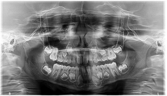
Bitewing radiographs revealed no caries and panoramic radiographs showed anodontia of permanent maxillary and mandibular right and left lateral incisors and maxillary right and left second premolars (Figure 3).

|
Figure 3 Panoramic radiograph showing the anodontia of permanent maxillary and mandibular right and left lateral incisors and maxillary right and left second premolars. |
The mother signed an informed consent form. The initial planned treatment consisted of dental prophylaxis of the upper and lower arches and aesthetic recontour of the anterior teeth to give the boy a better aesthetic and harmonized smile. The recontour was done with direct restorations in composite resin (Figure 4). After the conclusion of the restorative treatment the child was referred to orthodontic evaluation.
Discussion
The occurrence of chromosomal alterations largely consists of sporadic events during the replication of genes. Due to the imbalances, the amount and duplicate region lead to an unsure genotype/phenotype effect. The scarcity of reports in the literature comes from the fact that within a chromosome, the occurrence of the same duplication becomes a very rare event. The literature reports only 4 cases of trisomy of the long arm of chromosome 1 in part 1q44.1-6
The 1q duplications are considered rare genetic diseases, elucidated by the principle of extra copying of genetic material in one of the 46 chromosomes.1 The duplicated region within a chromosome ranges from the centromere (middle of the chromosome) to proximal and distal regions.1,2 Distal duplications are considered the most common, but still poorly reported; they comprise 1q31 to base 1q44 areas called pure trisomy or unbalanced translocations.6
Partial trisomy of chromosome 1 is a cytogenetic anomaly, and its phenotype is little-known.4 In most cases, unbalanced chromosomal translocations occur, making it difficult to associate signs and symptoms among patients. The damage caused to the patient is related to the duplication size and/or the appearance of a chromosome participant in the same region.5,6 Bartsch et al.4 state that more subtle alterations in distal regions seem to be associated with mental retardation or psychiatric illness, not showing dysmorphic characteristics, or presenting physical abnormalities. The authors suggest that the signs and symptoms depend on the size of the duplication, although in the reported cases the symptoms tended to increase according to the region that suffered the duplication; the closer to the center, the greater the damage. Main symptoms of 1q multiplication syndrome include mild to severe mental retardation, compromised pre and postnatal development, neuropsychomotor problems, relative macrocephaly, triangular face, prominent forehead, broad nasal bridge, retrognathia, flap ears, down-tilted palpebral fissures, micrognathia, ear and cardiac abnormalities.5,6 Thus, if these signs are suspected in early childhood in patients who may also present malformations in the hands and feet, the pediatrician may request genetic analysis to detect chromosomal abnormalities.4
Genetic tests are based on mapping through human DNA to detect alterations and duplications of chromosomal segments. Karyotype with a genetic band (G-band) consists of a collection of peripheral blood. It is a limited technique, and it can detect from 400-550 numeric or duplicate bands.4 The SNP-array technique used in the present case has the purpose of analyzing chromosomes with a higher resolution than the G-Band karyotype method, aiming to seek alterations that may explain the clinical signs and syntomes of patients submitted to this examination. This technique is considered one of the most sensitive for the identification of losses (deletions) and gains (duplications) of unbalanced submicroscopic chromosomal segments, through the general analysis of the genome, in a single experiment.6
The SNP-array is a powerful diagnostic tool for chromosomal abnormalities that can be used to complete the g-band karyotype analysis. However, traditional analysis techniques such as G-band karyotype analysis and FISH are still important, as the analysis of DNA microarrays cannot detect a translocation without a change in the number of copies or a region where no probe has been designed.1-6
It is interesting to note that due to the rarity of the Trisomy Syndrome of chromosome 1 and the scarce literature, the authors who reported it, performed a clinical evaluation, seeking through a detailed anamnesis, to point out problems that could have occurred from the gestational period to birth, together with research on family history to associate genetic inheritance.3 Only in one of the cited cases, genetic counseling was performed in parents and siblings, aiming to discover some chromosomal microdeletion. This is important because when it occurs in one of the parents, the genetic inheritance acquired by a child is possible.1,2 When both parents are carriers, the child, as in this case, can inherit part of this duplication. Thus, successively, genetic inheritance from chromosomal duplicated parents causes inheritance for future children, if they are unaware of this duplication.1-3
The present study is the first in the literature to present dental issues in a patient with trisomy of the long arm of chromosome 1 in region 1q44. The presence of diastema and alterations in the dental shape that caused great aesthetic disorder to the patient was corrected with the execution of direct restorations in composite resin. The direct restorative technique was chosen because it presented advantages such as reduced cost, preservation of the dental structure, speed of execution, and if necessary, it can be easily changed. This treatment proved to be efficient to return the patient´s self-esteem and provide resocialization, for the patient’s school social life was minimal, due to the bullying caused by his aesthetic appearance. Before the definitive restorative treatment of the anterior teeth, he always questioned the appearance of his teeth and compared his smile to those of his classmates. Afterwards, his mother noticed an important improvement in his socialization ability, and had her son smiling and talking happily again. Thus, not only his aesthetic problem was solved, but it also had a positive social impact, with great improvement to the boy’s quality of life.
Conclusion
Trisomy of the long arm of chromosome 1 is a very rare condition, with extremely limited literature available. In the dental area, the present study was the first case report to address dental issues expressed in a patient with this syndrome. It clarifies the main clinical characteristics and draws the attention of health professionals to the need for genetic evaluation, in the presence of these possible abnormalities. The improvement reported in the child's self-esteem and social interaction reinforces the impact of dentistry on the approach of functional and aesthetic treatments, upgrading his appearance and his function and social nature.
Acknowledgements
None.
Conflicts of Interest
None.
References
- National Institutes of Health. Chromosome 1q21.1 duplication syndrome. https://rarediseases.info.nih.gov/diseases/10591/chromosome-1q211-duplication-syndrome”. 2020.
- 1q21.1 microduplications. Understanding rare chromosome and gene disorders. https://rarechromo.org/media/information/Chromosome%20%201/1q21.1%20microduplications%20FTNW.pdf”. 2020.
- Coccé MC, Villa O, Obregon MG, et al. Duplication dup(1)(q41q44) defined by fluorescence in situ hybridization: delineation of the 'trisomy 1q42-->qter syndrome'. Cytogenet Genome Res. 2007;118(1):84‒86.
- Bartsch C, Aslan M, Köhler J, et al. Duplication dup(1)(q32q44) detected by comparative genomic hybridization (CGH): further delineation of trisomies 1q. Fetal Diagn Ther. 2001;16(5):265‒273.
- Watanabe S, Shimizu K, Ohashi H, et al. Detailed analysis of 26 cases of 1q partial duplication/triplication syndrome. Am J Med Genet A. 2016;170(A):908‒917.
- Morris M, Baroneza J, Teixeira P, et al. Partial 1q Duplications and Associated Phenotype. Mol Syndromol. 2015;6(6):297‒303.

